Africa Cancer Institute launch - 18 November 2013
Photos: Ronel Bester
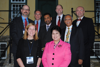
Group photo: (back left to right) Dr Oliver Bogler (University of Texas MD Anderson Cancer Center Senior Vice President Academic Affairs), Profs Eugene Cloete (SU Vice-Rector: Research & Innovation), Vikash Sewram (Director: ACI), Nico Gey van Pittius (SU Medicine & Health Sciences Deputy Dean: Research); Jimmy Volmink (SU Medicine & Health Sciences Dean), Prof Branislav Jeremic (Head: SU Division Radation Oncology). (front left) Dr Kathleen Schmeler (MD Anderson Cancer Center Assistant Professor, Department Gynaecology Oncology & Reproductive Medicine); Dr Shubhra Ghosh (MD Anderson Cancer Center Project Director of Global Academic Programs).
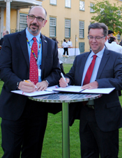
Signing of MOU: (left) Dr Oliver Bogler (University of Texas MD Anderson Cancer Center Senior Vice President Academic Affairs); Profs Eugene Cloete (SU Vice-Rector: Research & Innovation)
Photos: Lize Esterhuizen
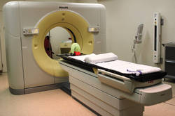 Big Bore CT scanner
» Click here to download photo
Four dimensional computer tomography (4D CT) represents a new and exciting step in cancer imaging and treatment planning. It takes images that not only capture the location of the tumor, but also capture its movement and the movement of the body's organs over time. This is very valuable for accurately treating tumors located on or near organs that move, such as those in the chest and abdomen.
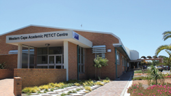 Western Cape Academic PET/CT Centre
» Click here to download photo
The Western Cape Academic PET/CT Centre gives patients in the public health sector in the Western Cape access to one of the best techniques in the world for early cancer diagnoses and management.
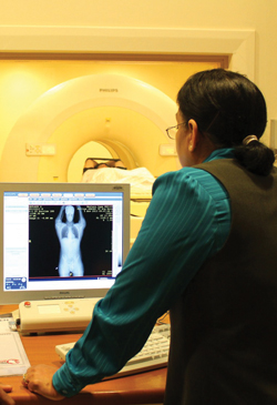 PET/CT scanner
» Click here to download photo
Radiographer scanning a PET/CT patient
A PET/CT scan combines a positron emission tomography (PET) scan with a computed tomography (CT) scan. In this way functional imaging (PET) using a small amount of radioactivity is combined with anatomical imaging (CT) to identify and localise abnormal processes in the body.
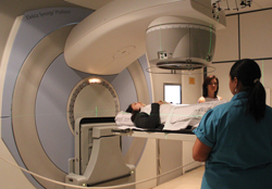 Elekta Linear accelerator
» Click here to download photo
Researchers and clinicians have access to high quality radiation technology equipment such as the Elekta Linear accelerator.
