Facilities
Undergraduate Microscopy Laboratory
The undergraduate microscopy laboratory is equipped with 142 state-of-the-art NIKON microscopes for teaching and enhancing the knowledge and skills of students in Cytology, Biology, and Histology. Slides are demonstrated on monitors before students can observe and draw “what they see" from their slides under the microscopes.
For more information, contact:
Mrs. Jodie Lemphane
Email: jilayman@sun.ac.za
Advanced Analytical Microscopy Facility
High-quality micrographs, ready for publication, can be taken with modern photo-imaging and analyzing compound and stereo microscopy systems. In addition, various services and training sessions are available and can be booked online.
For more information, booking details, and use of the microscopy systems, contact:
Mr. Jeffrey Pieterse
Email: jjp1@sun.ac.za
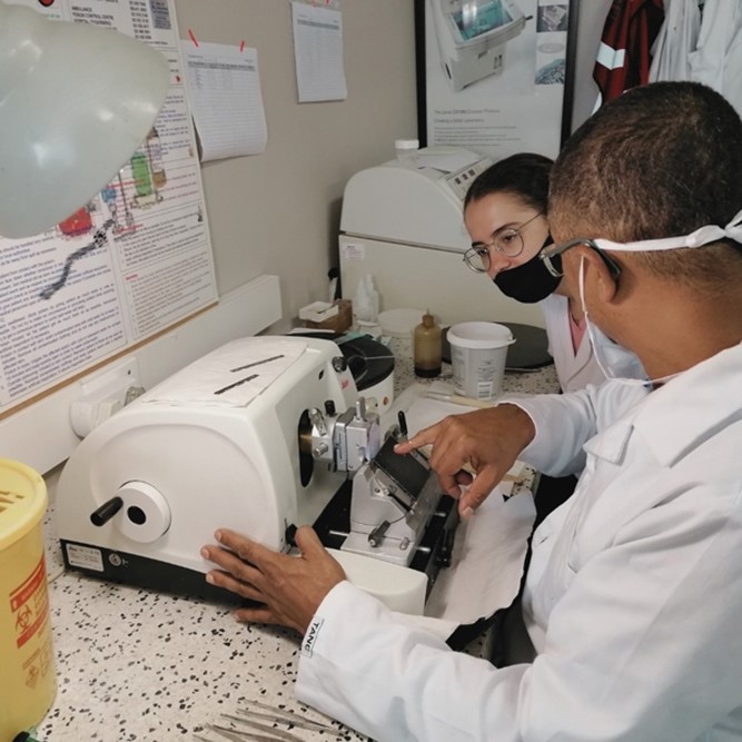
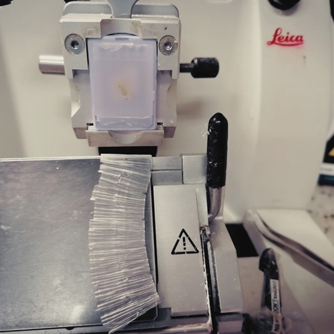
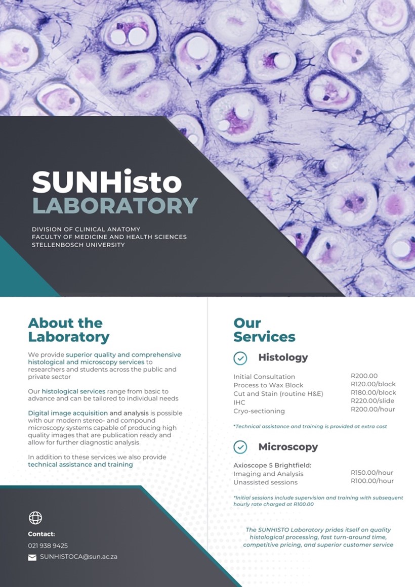
SUNHisto Laboratory
SUNHisto provides a comprehensive service of tissue analysis, including routine histological processes (paraffin and frozen sections for light-microscopy) and directed special staining (inclusive of ICC or IHC) for histological and pathological studies.
In addition to supporting high-tech research, our facilities and infrastructure are utilized by researchers from various departments such as Biochemistry, Physiology, Dermatology, Obstetrics and Gynaecology, MRC, and other academic units at Stellenbosch University. Furthermore, SUNHisto collaborates with other universities and private corporations, offering contract and outsourced research services. Consequently, the advanced academic expertise and infrastructure at SUNHisto benefit a wide range of researchers, the community, and the private sector.
For more information, contact:
Mr. Vuyo Mbovane (Histology Laboratory Manager)
Email: vuyom@sun.ac.za
Mr. Reggie Williams (Histology Laboratory Supervisor)
Email: rwilliams@sun.ac.za
Dissection Halls
The division houses four dissection halls in the BMRI South Building, with two halls located on the 4th floor and two on the 3rd floor. These halls are equipped with state-of-the-art dissection tables, an advanced extraction system, and a comprehensive collection of fully articulated skeletons and osteological materials. Additionally, a Macro-view projection system is available. Full-body Lodox® Statscan® images further enhance medical students' learning experience by aiding in the study of practical surface and radiologic anatomy.
For more information, contact:
Prof. Nanette Briers
Email: briers@sun.ac.za
Mr. Paul Pretorius
Email: pnbp@sun.ac.za
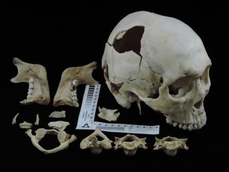
Stellenbosch Bone Repository
The division maintains an extensive repository of human skeletons obtained through the Body Donation Programme at the Division of Clinical Anatomy, Faculty of Medicine and Health Sciences, Tygerberg campus. This repository is utilized for educational and research purposes, in accordance with the National Health Act, Act 61 of 2003.
Access to the repository is restricted and requires approval. Prospective users must submit an application to the Division of Clinical Anatomy's Research Committee, consisting of the Stellenbosch Bone Repository research application form, a 2-page research proposal, and the CVs of the principal investigator and all co-investigators. Data collection can commence only after the Committee approves the study and receives proof of ethical approval from the applicant's home institution.
For more information, contact:
Dr. Mandi Alblas
Email: aa2@sun.ac.za
Mr. Keegan Meiring
Email: meiringk@sun.ac.za
Victim Identification at Stellenbosch University – ViSUN Unit
Forensic anthropology involves the utilization of biological anthropology principles within legal proceedings. The Victim Identification Service at Stellenbosch University (VISUN) collaborates with both the Victim Identification Centre (VIC) of the South African Police Services (SAPS) and the Forensic Pathology Services (FPS) of the Western Cape, offering support in the potential identification of unidentified skeletal, decomposed, or burnt human remains, particularly when conventional identifiers like DNA and fingerprinting are unavailable.
This service entails the provision of a comprehensive skeletal processing, analysis, and report submission, encompassing vital information such as the biological profile of the deceased, including sex, age-at-death, ancestry, and stature, as well as assessments of trauma and pathology. Additionally, a 3D surface scan of the skull is conducted to facilitate facial reconstruction efforts.
For more information, contact:
Dr. Mandi Alblas
Email: aa2@sun.ac.za
Equipment
Artec3D LEO Scanner
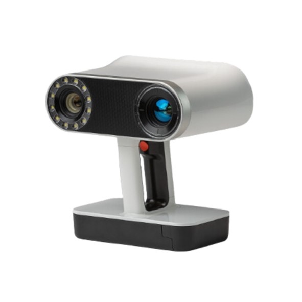
The Artec3D LEO Scanner is a cutting-edge 3D scanning device designed for rapid and highly accurate digitalization of objects. Equipped with advanced features, the LEO Scanner captures intricate details with high precision, making it an invaluable tool for anatomical research and education.
The Artec3D LEO Scanner is a vital addition to our division's array of high-tech equipment, supporting both educational and research endeavors in the field of clinical anatomy.
For more information, contact:
Ms. Janine Correia
Email: jcorreia@sun.ac.za
Dr. Nadine Rampf
Email: nrampf@sun.ac.za
Zortrax M300 Dual 3D printer
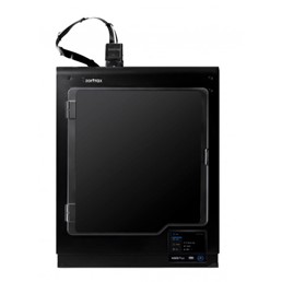 The Zortrax M300 Dual 3D Printer is a state-of-the-art additive manufacturing device designed for creating highly detailed and durable 3D models. This advanced printer is an essential tool for both educational and research purposes in the Division of Clinical Anatomy, enabling the production of precise anatomical models and prototypes.
The Zortrax M300 Dual 3D Printer significantly enhances our division's capabilities, facilitating the creation of detailed anatomical models that aid in the visualization and understanding of complex structures, thereby enriching both teaching and research activities in clinical anatomy.
For more information, contact:
Ms. Janine Correia
Email: jcorreia@sun.ac.za
Dr. Nadine Rampf
Email: nrampf@sun.ac.za
Ultrasound
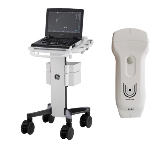 Ultrasound (US) is increasingly utilized across various medical specialties as a diagnostic tool, prompting medical faculties to incorporate imaging more extensively into their curricula. Integrating US into undergraduate instruction offers numerous benefits. It can enhance anatomical knowledge and improve the visual understanding of anatomy. The cost-effectiveness and portability of US make it a valuable addition to traditional anatomy teaching methods and it is currently being implemented in undergraduate programmes in the Division of Clinical Anatomy. Additionally, US is employed in postgraduate research studies within the division.
For more information, contact:
Ms. Janine Correia
Email: jcorreia@sun.ac.za
Versana Active Ultrasound System and the U-Image Point of Care
Ultrasound
BrainBit EEG Headbands
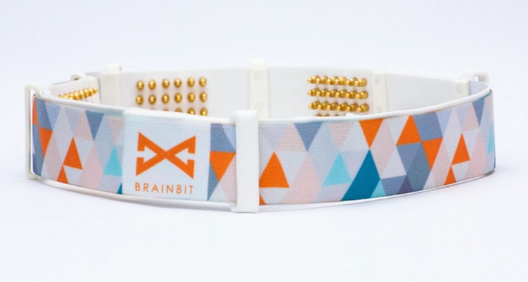 The BrainBit EEG Headbands are innovative wearable devices designed to measure and record brainwave activity. These headbands are integral to our division's efforts in anatomical sciences education and sleep research, providing real-time insights into neurological functions and sleep patterns.
The BrainBit EEG Headbands are a crucial part of our equipment repertoire, advancing our capabilities in both educational and research domains. By providing detailed insights into brainwave activity, these headbands enrich our understanding of neurological functions and sleep dynamics, thereby supporting our mission to excel in anatomical sciences education and sleep research.
For more information, contact:
Ms. Janine Correia
Email: jcorreia@sun.ac.za
Dr. Nadine Rampf
Email: nrampf@sun.ac.za
