Equipment database Light Microscopy
Zeiss LSM780 confocal microscope with ELYRA PS1 super-resolution platform
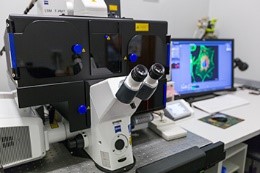
Location: Mike de Vries Building, Merrimen Avenue, Room 2024
Function and Purpose:
The LSM780 ELYRA PS1 microscope is a combined confocal and super-resolution microscope. The instrument features 6 laser lines for confocal acquisition and with a 32 channel GaAsP detector, it can acquire fluorescence signal from all commercial fluorescence markers from the violet to the far red range. The microscope allows for multi-colour imaging, FRET (Foerster Resonance Energy Transfer), Bleaching experiments, Z-sectioning and co-localisation. With an on-stage incubator and definite focus feature it is also suitable for live cell studies to follow dynamic processed.
The microscope is equipped with two different super-resolution modalities, including structured illumination microscopy (SR SIM) and single molecule localisation mapping (SMLM), previously known as PALM/STORM. This allows for improved resolution down to 100 nm and 30 nm for the two modalities respectively. This enables researchers to study samples even down to protein level.
Carl Zeiss Axio Petrographic Light Microscope
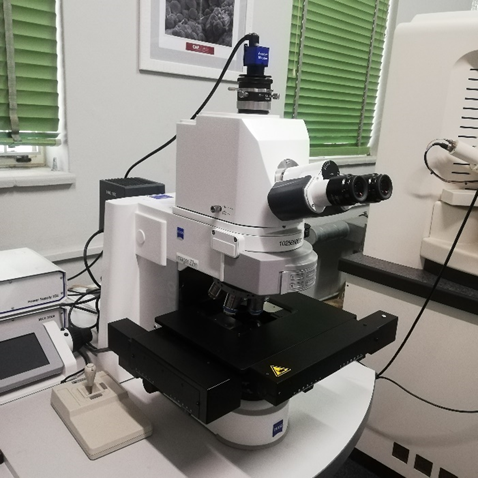
Location: Chamber of Mines Building, Cnr Ryneveld & Merriman Street, Room 1034/5
Function and Purpose:
It is fully correlated with the MERLIN FE SEM by the Carl Zeiss Shuttle & Find correlative microscopy interface for light- and electron microscopes.
The CLEM platform capabilities (designed for correlative light and electron microscopy-CLEM by Carl Zeiss), allows the use of the sample in both the Elyra 780 system as well as the applied for FE SEM system, so that the sample is stably fixed in both instruments during the acquisition process. This holder contains three coordinate markers which define a coordinate system that are calibrated in the Shuttle & Find module. This software module can operate intrinsically in the ZEN 2012 software or can operate separately as an independent module on both the FE SEM or Elyra system.


ZEISS SteREO Light Microscope with Axio Software
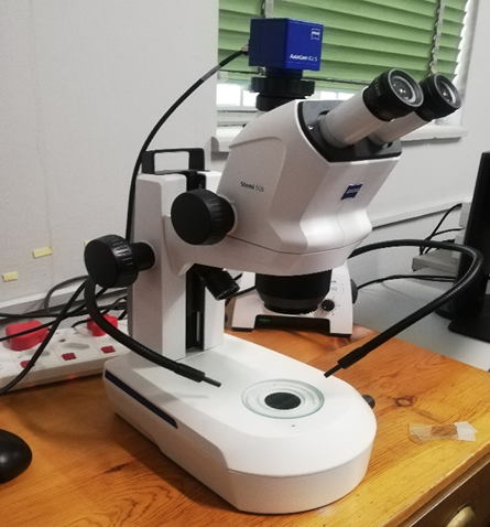
Location: Chamber of Mines Building, Cnr Ryneveld & Merriman Street, Room 1034/5
Function and Purpose:
Light microscopy is used to quickly observe and record sample features at lower magnifications. The light microscopy images with reveals unrestricted depth of focus, or even 3D images of sample features that can be viewed from different angles. The most obvious attribute of the light microscope is its ability to bridge the magnification range required for basic inspection, imaging and documentation, at low magnifications with little sample preparation needed.
Tygerberg equipment
Zeiss Axio Observer 7 Inverted Microscope

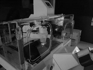
Location: Clinical Building, Room 3046
Function and Purpose:
The Zeiss Axio Observer 7 Inverted Microscope is a platform for either light and/or fluorescence microscopy imaging to analyse fixed cells or tissue sections. An added feature to this unit is an incubation chamber, offering the option of performing live cell imaging under control physiological conditions. The instrument delivers: Brightfield, Dark field, Phase contrast and DIC modalities, 7-fluorescent channels of detection and is set up with at 5x, 10x, 20x, 40x (water/glycerol) and 63x (oil immersion) objective.
