Equipment database Electron Microscopy
ZEISS MERLIN high resolution field emission scanning electron microscope (FE SEM)
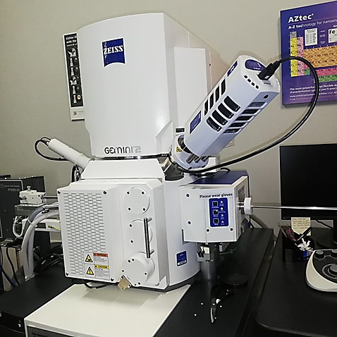
Location: Chamber of Mines Building, Cnr Ryneveld & Merriman Street, Room 1034/5
Function and purpose:

The ZEISS Merlin FE SEM has nano-scale image and micro-and cryo-EDS analytical capabilities. The MERLIN combines ultra-fast analytics and high resolution imaging and is capable of a wide range of beam flexibility, from a voltage of 20V to 30kV and beam currents of 7pA to 40 nA, as well as resolutions of up to 0.6 nanometers at 30kV and 1.6 nanometers at 1kV.
The system is fitted with a number of detectors for imaging, including: an in-lens and chamber secondary (SE) detectors (nano & micron-scale imaging), an in-column energy selective backscattered (ESB) electron detector, a retractable 5 diode backscattered electron detector (BSD) (nano-BSE & 5-diode detector nano to micron imaging), a cathodoluminescence (CL) detector and a scanning transmission electron microscopy (STEM) detector with a resolution of 0.6 nanometer.
EDX (Elemental phase mapping, linescans, spot, cryo, feature & automate analysis)
Analytical capability is provided by the Oxford Instruments XMax 20mm2 detector for high resolution spectra and high spatial resolution maps using energy dispersive X-ray spectrometry (EDS). Furthermore, the instrument has a Quorum cryostage for micro-quantitative analysis of beam sensitive samples, as well as a facility for local charge compensation (CC) for the analysis of insulating samples.


ZEISS EVO® MA15 EDS&WDS SEM
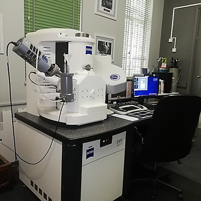
Location: Chamber of Mines Building, Cnr Ryneveld & Merriman Street, Room 1034/5
Function and purpose:
The Zeiss EVO® MA15 EDS&WDS SEM is fitted imaging detectors, including secondary detectors (SE) and backscattered electron detector (BSD). Analytical capability is provided by the Oxford Instruments XMax 20mm2 Electron Dispersive X-ray (EDS) detector and Wave Dispersive X-ray Spectrometer (WDS) for analytical capabilities widely applied elemental microanalysis method capable of identifying and quantifying all elements in the periodic table except H, He, and Li. By following the “k‐ratio" (unknown/standard) measurement protocol development for electron‐EDS/WDS. EDS&WDS is a non-destructive analysis technique used to obtain elemental information about a range of materials by measuring characteristic x-rays within a small wavelength range. EDS/WDS analytical techniques include the following-: elemental phase mapping, linescans/traverse, spot/area, feature & automate analysis.
Instruments at Tygerberg:
Thermo Fisher Apreo 3D FEG SEM

Location: Clinical Building, 7th floor, Room 7063
Function and purpose:
Unraveling complex 3D architecture of cells and tissues in their natural context is crucial for the structure function correlation in biological systems. In recent years, there have been considerable advances in SEM-based methods for 3D reconstruction of large tissue volumes. Serial Block-Face SEM (SBF-SEM) combines in situ sectioning and imaging of plastic embedded tissue blocks within the SEM vacuum chamber in a fully automated fashion for reconstruction of large tissue volumes. Until now, the axial resolution was limited by the minimal section thickness that can be cut from the block-face; however, with a combination of SBF-SEM and Multi-Energy Deconvolution SEM (MED-SEM), the Thermo Scientific™ Apreo™ VS now enables large-volume imaging with truly isotropic 3D resolution.

