Vibrational Spectroscopy
| 
|
The CAF Vibrational Spectroscopy Unit at Stellenbosch University in the Western Cape houses the hyperspectral imaging equipment which can be used for assessing the chemical, physical or biological differences of an organic sample.
Spectroscopy is the study of how electromagnetic radiation interacts with matter. Hence, infrared spectroscopy is focussed on the response to the infrared spectral region of the electromagnetic spectrum. This region can be divided into the near- (780 – 2500 nm), mid- (2500 – 25 000 nm) and far infrared (25 000 – 1 000 000 nm) regions.
The instruments available in the Vibrational spectroscopy unit function in the visible (400 – 700 nm) and near infrared (NIR) region. Radiation, here referred to as 'light', in the NIR region typically excite the vibration of the major X-H bonds such as C-H, O-H and N-H, which are common constituents of organic molecules. During spectroscopy, the incident light interacts with these molecules and different molecules are sensitive to different wavelengths. The recorded spectra indicate how the incident light was absorbed or scattered by the sample allowing information such as the physical, chemical or biological differences of the sample and its constituents to be collected.
In conventional NIR spectroscopy a small portion of the sample is scanned, and an average spectrum or prediction value is determined and taken to be representative of the entire sample. However, this information may not be a true reflection of the 'non-scanned regions' of the sample especially if the distribution of the property is heterogenous. Hyperspectral imaging (HSI) addresses this limitation of conventional spectroscopy.
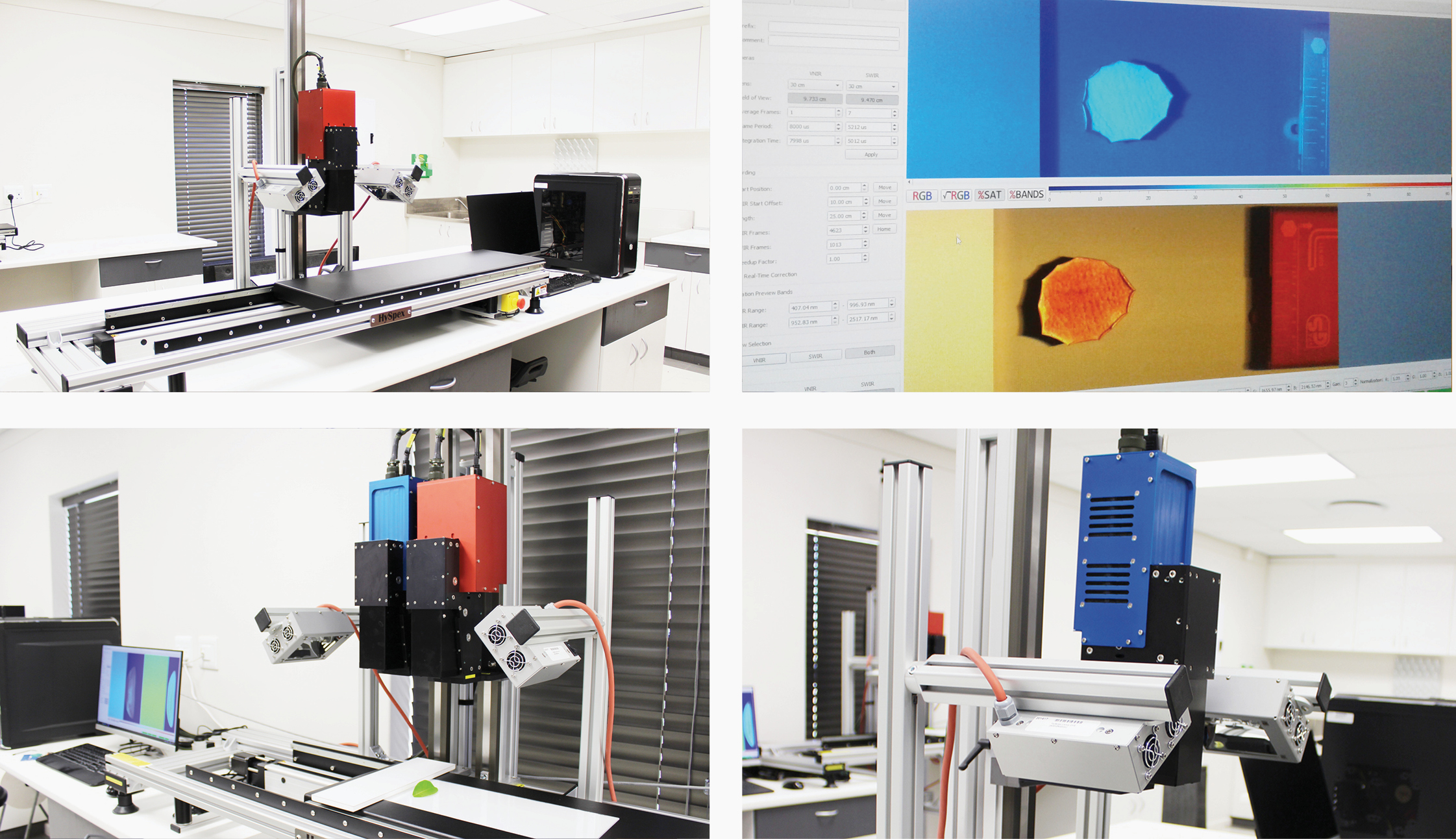
Hyperspectral imaging combines digital imaging with spectroscopy. In contrast to conventional NIR spectroscopy, the entire sample is analysed and imaged. The HSI camera records the contiguous spectral information for up to a few hundred spectral bands for each pixel in the image. The hyperspectral image which is formed is therefore made up of a stack of several hundreds of 2-D spatial images of the same sample. Each layer represents a narrow wavelength band and by stacking the layers, a complete spectrum (eg. 700 – 1000 nm) for each pixel is formed. The collection of spectral layers is known as the hypercube. This unique spectral signature or fingerprint of each pixel in the images facilitates the detection of subtle differences in spectra between pixels. Study of these spectral differences enables various components to be investigated simultaneously and can be used to resolve the chemical composition and distribution, and the physical properties of the sample.
Since hyperspectral images contains a large data set, it can be challenging to analyse and extract meaningful information. The application of chemometrics allows the reduction of the data set whilst retaining important spectral information and identifying important characteristics of an image. Multivariate image analysis therefore enables the properties of the sample under investigation to be correlated to the hyperspectral image.
The advantage of using HSI, is that this technique is fast, accurate and allows samples to be analysed non-destructively and non-invasively with minimum or no sample preparation. This technology has applications across a broad spectrum of fields with applications reported for food quality and safety, medical, agricultural, archaeological, palaeontological and the pharmaceutical industries.
The unit which is located in the Food Sciences Department is equipped with two systems; a Visible Near Infrared (VNIR) and Short Wave Infrared (SWIR) imaging system operating in the 400 – 1000 nm and the 950 – 2500 nm spectral range, respectively. Users have the option to image samples in the reflectance or transmission mode. Each set-up have its own LabRack with sample moving stage. A variety of lenses are also available for use. Four dedicated computers with the Evince and Breeze software is available for acquiring images and for data analysis and processing.
Clients from both academia and industry are welcome to make use of the available equipment in this unit.
Equipment available in the unit
Further reading
Manley M (2014). Near-infrared spectroscopy and hyperspectral imaging: non-destructive analysis of biological materials. Chemical Society Reviews 43: 8200
Sendin, K., Williams, P.J. & Manley, M. (2016). Near infrared hyperspectral imaging in quality and safety evaluation of cereals. Critical Reviews in Food Science and Nutrition, 1-16.
This information will be updated regularly - please visit again.
