|
|
The
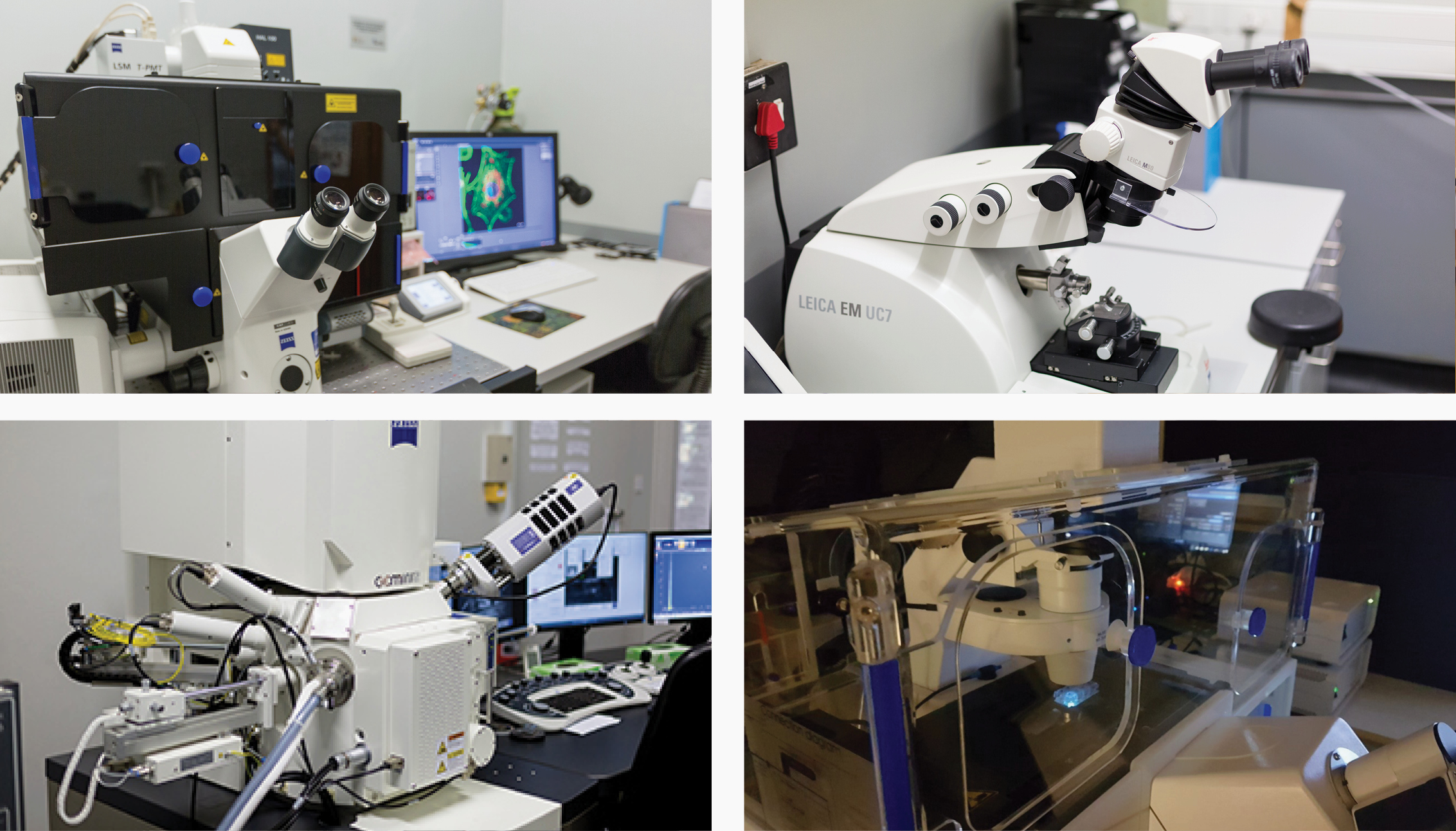
Microscopy Unit provides access to advanced light and electron microscopes as well as the expertise of dedicated imaging scientists. We support clients from both research and industry, and our applications range from material science to biomedical research.
Hence, we provide custom image analysis, comprehensive sample preparation for both light and electron microscopy, and consultation.
Lize Engelbrecht (CAF Microscopy Unit) attended a Global BioImaging Conference in Uruguay last month. See the video where an interview with Lize is included.
|
|
| Serial Block-Face SEM
Sample: Zebrafish Larvae Brain
Instrument: ThermoFisher Apreo Volumescope
Description: Serial Block-Face SEM allows for volumetric acquisition of resin embedded samples in which structures of interest can be segmented and assessed in 3D. In this example, the volumetric rendering of connected neurons in the cortical region of a zebrafish larvae brain is shown.
| Live Cell Imaging
Sample: Glioma Cells
Instrument: Zeiss LSM 780
Description: Our temperature and gas control stage allows for live cell imaging of cellular and organellar events. Here, parts of the mitochondrial were photoactivated in order to quantify the rate at which they fuse and divide.
|
CONTACT
Lize Engelbrecht | Madelaine Frazenburg |
Janica Theron
Physical Address
Stellenbosch University, Room 2022, Mike de Vries Building, Merriman Avenue, STELLENBOSCH, 7600
Stellenbosch University, Room 1034/5, Chamber of Mines Building, Cnr Ryneveld & Merriman Street, STELLENBOSCH, 7600
Tygerberg Medical Campus, BMRI Building, Room 2106, BELLVILLE, 7493
Postal Address
Stellenbosch University, Private Bag X1, Matieland, STELLENBOSCH, 7602
