​​​​​NuMeRI Node for Infection Imaging​ 

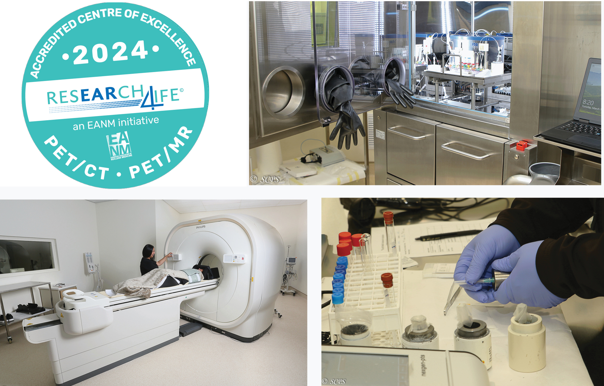
EARL accredited PET/CT Centre of Excellence​

> ​REGIST​​ER FOR A C​​​A​​​​F​​ ID
​> RE​GISTER TO MA​KE A ​​BOOKING
> ​MAKE A BOOKIN​G​​​ 
​> E​​QUIPMENT GALLERY​​​​​
​> SCAN PREPARATION
​> RESEARCH CLIENTS
> ​PRIVATE C​LIENTS
​> PACS
​> CON​TAC​​T​ 

​​​​


​​The NuMeRI Node for Infection Imaging​ in the Western Cape is a state of the art PET/CT research facility, and a core node of the Nuclear Medicine Research Infrastructure (NuMeRI), which forms part of the South African Research Infrastructure Roadmap (SARIR). The N​ode is hosted by the Central Analytical Facilities (Stellenbosch University) and is located at Tygerberg Hospital, in Cape Town.

​Introduction

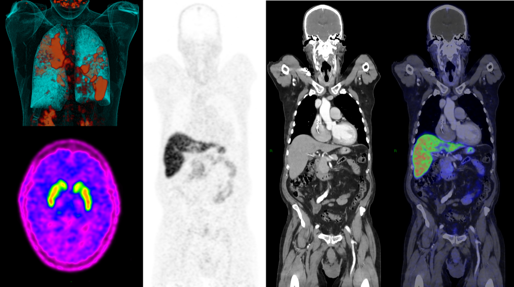
Nuclear imaging exploits a fundamental property of certain unstable atoms, namely that they will undergo radioactive decay by either directly or indirectly emitting gamma photons. By labelling biological molecules of interest with positron-emitting radionuclides it becomes possible, using specialised equipment, to image the position of decay events and hence, the three-dimensional location of the molecule in question. This imaging is performed with a positron-emission tomography (PET) scanner which in modern systems is routinely combined with a conventional (x-ray) computed tomography (CT) system. The combined ability of such PET/CT systems to quantitatively and dynamically image the behaviour of radiolabelled molecules (radiotracers) with a high degree of accuracy, and to localise their distribution precisely with the anatomical definition provided by their CT component, has revolutionised biological research.

​​Our Equipment

The keystone of the NII is its Philips Vereos digital PET/CT scanner. Due to replacement of traditional photomultiplier tubes with solid-state silicon photomultipliers, it is possible for the Vereos PET to achieve 1:1 photon counting, leading to unprecedented accuracy and effective sensitivity. The system is integrated with a Philips Ingenuity 64-channel (128-slice equivalent) CT with iDose4. The latest-generation iterative reconstruction capability of iDose4 allows for low-dose diagnostic CT scanning with superior image quality to conventional FBP-based reconstruction techniques. The combination of high quantitative accuracy, superb effective sensitivity, and low-dose/high-quality CT scanning makes the Vereos the ideal nuclear imaging tool for research applications.

Supporting our PET/CT scanner is an advanced radiopharmacy. This unit boasts a dispensing laboratory, a hot laboratory, a clean room, and a quality control laboratory. In these facilities, our staff are able to perform stringent quality control of injected radiopharmaceuticals, to label kit-based radiotracers, and to perform research and de-novo syntheses of radiolabelled molecules of interest.

​Our Staff

The Unit has 7 permanent staff members, including a nuclear medicine staff scientist, a radiopharmacist, three nuclear medicine radiographers, a practice manager, and an administrative assistant.​ This team offers researchers the support they need to perform the molecular imaging and analysis they require. They are supported by academic staff of the Divisions of Nuclear Medicine and Radiodiagnosis of the Faculty of Medicine and Health Sciences of Stellenbosch University, who have a long track-record in PET/CT research.

​Studies offered

The NII currently offers routine scanning with four different radiotracers:

  • F-18 fluorodeoxyglucose (FDG): a radioactive glucose analogue commonly used to image inflammatory processes; certain malignancies; regional brain metabolism; and cardiac viability.
  • F-18 fluoro-DOPA (FDOPA): a radioactive amino acid transporter commonly used to image integrity of the striatal dopaminergic system; brain tumours; and selected malignancies of the sympathetic nervous system.
  • Ga-68 PSMA: a radiotracer with high affinity for prostate cancer commonly used to stage high-risk cases and detect prostate cancer recurrence.
  • Ga-68 DOTANOC: a radioactive somatostatin analogue commonly used to image selected neuroendocrine tumours.

The NII also performs research with other radiotracers labelled on site. Interested parties should please contact the unit for further information.

Examples of PET/CT scans using different radiotracers appear below:


Limited field PET (using F-18 FDG) fused with serially acquired CT in a patient with tuberculosis. Intensity of uptake on the PET correlates with metabolic activity within lesions.

Anterior oblique projection of an F-18 FDOPA PET scan showing normal presynaptic dopaminergic function in bilateral striatal structures of the brain.


Whole body projection of a PET scan performed using Ga-68 prostate-specific membrane antigen (PSMA). In addition to normal biodistribution, spread of prostate cancer to multiple lymph nodes (arrows) is visible.PET images acquired using Ga-68 DOTANOC (a somatostatin analogue) fused with serially acquired CT, of a patient suspected of having a neuroendocrine tumour of the small bowel. This tumour (arrow) was undetectable on anatomical imaging or on endoscopy but was confirmed by histology.


​PET research at SU

A selection of original research articles with Stellenbosch University collaborators in which PET/CT was used appears below:

  • Burger C, Holness JL, Smit DP, Griffith-Richards S, Koegelenberg CFN, Ellmann A. The Role of 18F-FDG PET/CT in Suspected Intraocular Sarcoidosis and Tuberculosis. Ocul Immunol Inflamm. Published online November 19, 2019:1-7. doi: 10.1080/09273948.2019.1685109
  • Doruyter A, Dupont P, Taljaard L, Stein DJ, Lochner C, Warwick JM. Resting regional brain metabolism in social anxiety disorder and the effect of moclobemide therapy. Metab Brain Dis. 2018;33(2):569-581. doi: 10.1007/s11011-017-0145-7
  • Du Toit R, Shaw JA, Irusen EM, von Groote-Bidlingmaier F, Warwick JM, Koegelenberg CFN. The diagnostic accuracy of integrated positron emission tomography/computed tomography in the evaluation of pulmonary mass lesions in a tuberculosis-endemic area. S Afr Med J. 2015;105(12):1049-1052. doi: 10.7196/SAMJ.2015.v105i12.10300​
  • Esmail H, Lai RP, Lesosky M, et al. Characterization of progressive HIV-associated tuberculosis using 2-deoxy-2-[18F]fluoro-D-glucose positron emission and computed tomography. Nat Med. 2016;22(10):1090-1093. doi: 10.1038/nm.4161
  • Fitzgerald BL, Islam MN, Graham B, et al. Elucidation of a Human Urine Metabolite as a Seryl-Leucine Glycopeptide and as a Biomarker of Effective Anti-Tuberculosis Therapy. ACS Infect Dis. 2019;5(3):353-364. doi: 10.1021/acsinfecdis.8b00241
  • Kleynhans J, Rubow S, le Roux J, Marjanovic-Painter B, Zeevaart JR, Ebenhan T. Production of [68 Ga]Ga-PSMA: Comparing a manual kit-based method with a module-based automated synthesis approach. J Label Compd Radiopharm. 2020;63(13):553-563. doi:10.1002/jlcr.3879​​​
  • Le Roux J, Rubow S, Ebenhan T, Wagener C. An automated synthesis method for 68Ga-labelled ubiquicidin 29–41. J Radioanal Nucl Chem. 2020;323(1):105-116. doi: 10.1007/s10967-019-06910-1​
  • Le Roux J, Rubow S, Ebenhan T. A comparison of labelling characteristics of manual and automated synthesis methods for gallium-68 labelled ubiquicidin. Appl Radiat Isot Data Instrum Methods Use Agric Ind Med. 2021;168:109452. https://doi.org/10.1016/j.apradiso.2020.109452
  • ​Malherbe ST, Chen RY, Dupont P, et al. Quantitative 18F-FDG PET-CT scan characteristics correlate with tuberculosis treatment response. EJNMMI Res. 2020;10(1):8. doi: 10.1186/s13550-020-0591-9
  • Malherbe ST, Dupont P, Kant I, et al. A semi-automatic technique to quantify complex tuberculous lung lesions on 18F-fluorodeoxyglucose positron emission tomography/computerised tomography images. EJNMMI Res. 2018;8(1):55. doi: 10.1186/s13550-018-0411-7​
  • Malherbe ST, Shenai S, Ronacher K, et al. Persisting positron emission tomography lesion activity and Mycobacterium tuberculosis mRNA after tuberculosis cure. Nat Med. 2016;22(10):1094-1100. doi: 10.1038/nm.4177​
  • Morkel M, Ellmann A, Warwick J, Simonds H. Evaluating the Role of F-18 Fluorodeoxyglucose Positron Emission Tomography/Computed Tomography Scanning in the Staging of Patients With Stage IIIB Cervical Carcinoma and the Impact on Treatment Decisions. Int J Gynecol Cancer. 2018;28(2):379-384. doi: 10.1097/IGC.0000000000001174
  • ​Prince D, Rossouw D, Davids C, Rubow S. Development and Evaluation of User-Friendly Single Vial DOTA-Peptide Kit Formulations, Specifically Designed for Radiolabelling with 68Ga from a Tin Dioxide 68Ge/68Ga Generator. Mol Imaging Biol. 2017;19(6):817-824. doi: 10.1007/s11307-017-1077-7
  • Prince D, Rossouw D, Rubow S. Optimization of a Labeling and Kit Preparation Method for Ga-68 Labeled DOTATATE, Using Cation Exchange Resin Purified Ga-68 Eluates Obtained from a Tin Dioxide 68Ge/68Ga Generator. Mol Imaging Biol. 2018;20(6):1008-1014. doi: 10.1007/s11307-018-1195-x
  • Shaw JA, Irusen EM, Groote-Bidlingmaier F von, et al. Integrated positron emission tomography/computed tomography for evaluation of mediastinal lymph node staging of non-small-cell lung cancer in a tuberculosis endemic area: A 5-year prospective observational study. S Afr Med J. 2015;105(2). Accessed May 10, 2019. https://www.ajol.info/index.php/samj/article/view/114031
  • Simonds H, Botha MH, Ellmann A, et al. HIV status does not have an impact on positron emission tomography-computed tomography (PET-CT) findings or radiotherapy treatment recommendations in patients with locally advanced cervical cancer. Int J Gynecol Cancer. 2019;29(8):1252-1257. doi: 10.1136/ijgc-2019-000641
  • Thompson EG, Du Y, Malherbe ST, et al. Host blood RNA signatures predict the outcome of tuberculosis treatment. Tuberculosis. 2017;107:48-58. doi: 10.1016/j.tube.2017.08.004
  • Twycross SH, Burger H, Holness J. The utility of PET-CT in the staging and management of advanced and recurrent malignant melanoma. South Afr J Surg Suid-Afr Tydskr Vir Chir. 2019;57(3):44-49.
  • Vuletic D, Dupont P, Robertson F, Warwick J, Zeevaart JR, Stein DJ. Methamphetamine dependence with and without psychotic symptoms: A multi-modal brain imaging study. NeuroImage Clin. 2018;20:1157-1162. doi: 10.1016/j.nicl.2018.10.023