Stellenbosch Campus:
|
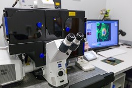
Zeiss LSM780 with ELYRA PS1 platforms
Location: Mike de Vries Building, Room 2022
- A high sensitivity, inverted, point-scanning confocal microscope
- Equipped with an on-stage incubator for CO2, humidity and temperature control for live cell imaging.
- Equipped with super-resolution platforms: SR SIM and PALM/STORM/SMLM
- Capable of Total Internal Reflection Fluorescence (TIRF)
- Lasers available:
- 405nm diode laser.
- Argon laser with 3 excitation lines (458nm, 488nm, 514nm)
- 561 nm laser
- 633 nm laser
|
|
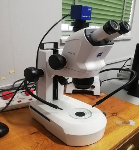
Zeiss Stemi 508 Stereo Microscope
Location: Chamber of Mines Building, Room 1034/5,
Two reflected light LED and one transmitted light LED Choose between mirror-based transillumination or the flat brightfield-darkfield transilluminator Camera: Zeiss AxioCam ICc 5 colour camera Magnification: 5:1 Software: ZEN 2 Core Imaging Software
|
|
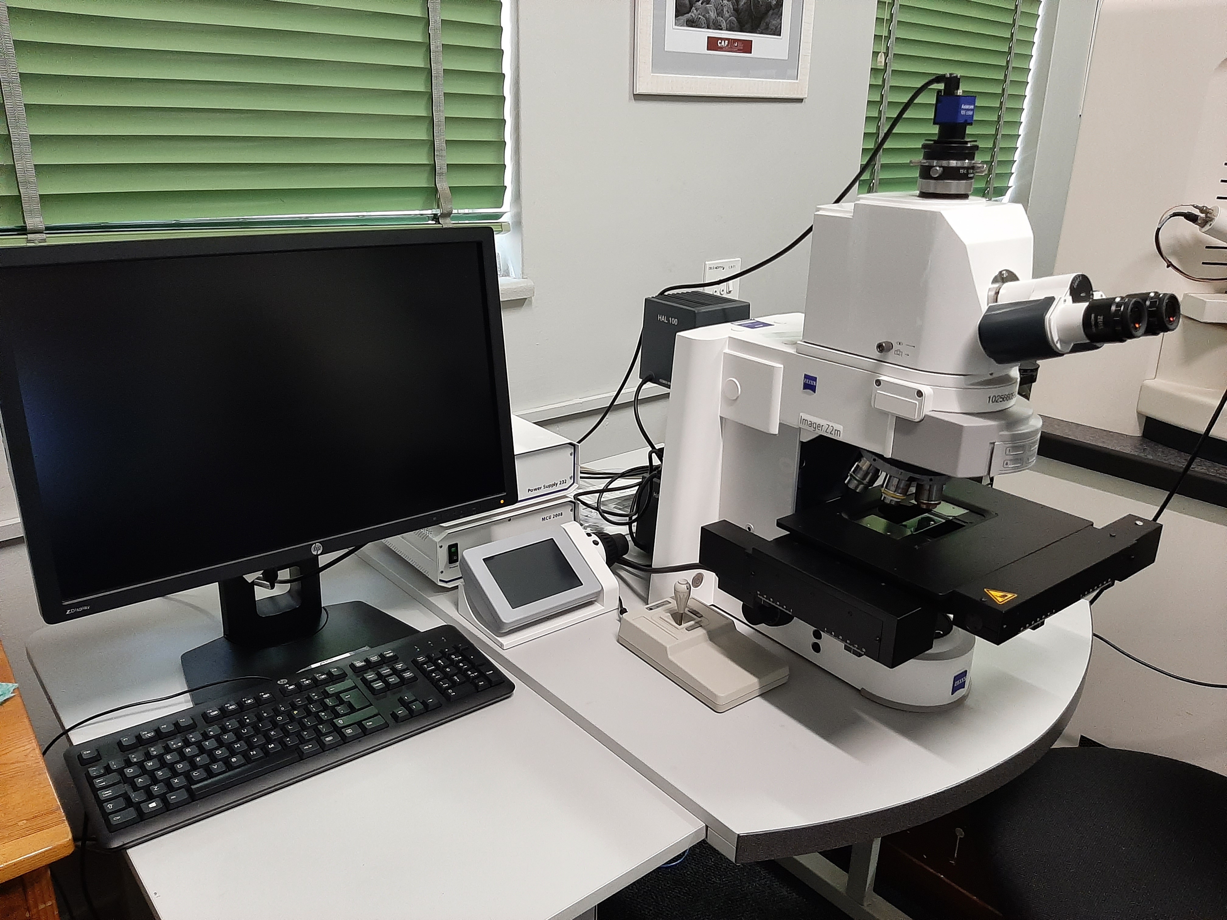
Zeiss AxioImager.Z2m petrographic microscope
Location: Chamber of Mines Building, Room 1034/5,
- Allows for brightfield, darkfield and polarized microscopy, as well as DIC
- Allows for correlative microscopy
- Software: Axio Vision SE64 Release 4.9.1
- Objectives: 20X, 10X, 2.5X, 20XEPI, 40X, 50X
- Camera: Zeiss AxioCam 105 color
|
Tygerberg Campus: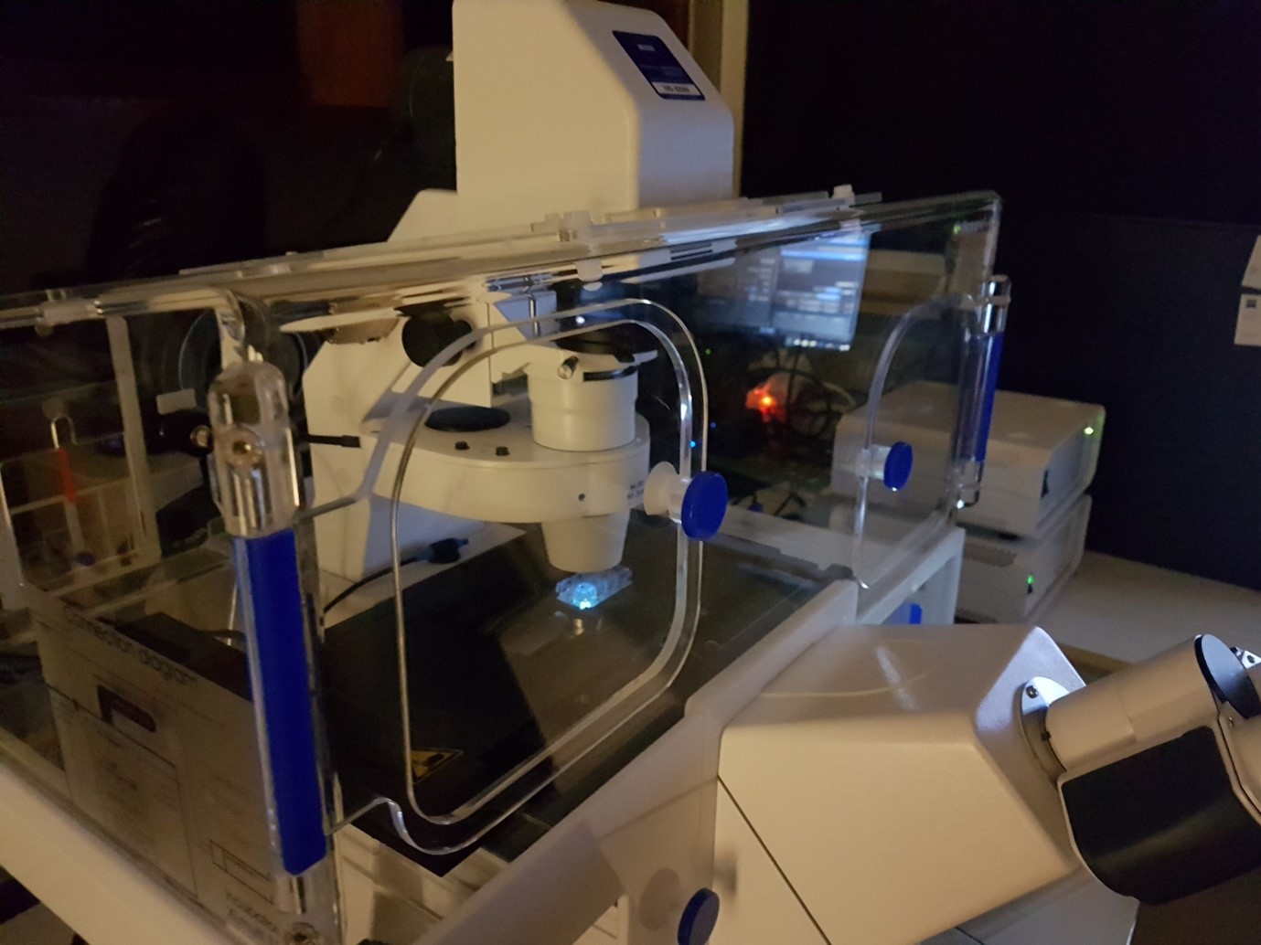
|
Zeiss AxioObserver 7 - Live-cell fluorescence microscope
Location: Clinical building, Room 7063 - Colibri LED light source for illumination with 7 wavelengths and filters to detect all the main fluorophores such as DAPI, Hoechst, GFP, Alexafluor 488, CFP, YFP, Texas Red, Alexafluor 647 and many more.
- Additional BP filters for FRET experiments using CFP and YFP
- Monochrome camera for fluorescence imaging and a colour camera for histological imaging
- Brightfield, Dark field, Phase contrast and DIC modalities
- 5x, 10x, 20x, 40x objectives for imaging through culture plates
- 40x (water/glycerol immersion) and 63x (oil immersion) for imaging through cover slips
- Live Imaging: Incubation chamber with settings to control temperature, CO2/O2 and humidity levels.
|
