In a first for Africa, a correlative nano-imaging platform has been established at Stellenbosch University's Central Analytical Facilities (CAF) with the acquisition of a new state-of-the-art electron microscope.
The new Zeiss MERLIN field emission gun scanning electron microscope can resolve features as small as 0.6 nano-meters in size, as well as measure the chemical composition of delicate glass, mineral and biological samples.
The new microscope has now been correlated with a Zeiss super-resolution scanning laser microscope which has recently been up-graded to achieve additional super-resolution capabilities.
The correlation of these two cutting edge microscopes has made possible the first nano-resolution correlative light (laser) and electron microscopy (CLEM) platform in Africa, explains Ms Lize Engelbrecht, manager of CAF's Fluorescent Microscopy Unit.
Dr Angelique Coetzer, staff analyst at CAF's Scanning Electron Microbeam Unit where the new electron microscope is housed, says scientists will now be able to both identify an area of concern on the nanoscale using fluorescence and further explore the structure of that feature in more detail using electron imaging. Samples can be transferred from one microscope system to another in just a few minutes. Furthermore, the search for the same nanometer-scale region of interest is fully automated.
Dr Coetzer says students and researchers from various disciplines, in particular from microbiology, physiological sciences, geology, engineering and polymer science, have already started to explore the exciting possibilities offered by the new technology.
There is also significant interest from industry in the chemical and image analysis capabilities of the new equipment and the nano-imaging platform. Clients are from the mineral and diamond exploration industry, nanofiber manufacturers, the food, wine and spirit industry, as well as the munitions, welding/metal, water filters, jewellery and paint industries.
The official launch of the new equipment, and a showcase of the first results from the correlative microscopy (CLEM) platform, will take place on Tuesday 11 October 2016.
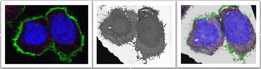
In the images above: Previously scientists were able to look into, for example, a cell by using fluorescent markers to identify contents of the cell which are of interest. In the fluorescence image above left, virus particles that entered cells, the cell membrane is stained green and the nucleus (which controls cellular function) is visible in blue. However, up till now it was impossible to obtain a clear image of the structure of the part of the cell that needs to be analysed. With the new Zeiss MERLIN field emission gun scanning electron microscope, the internal cellular structure of that part of the cell can now be visualised – see the two images (above middle and right) of the same area of the cell.
Technical information
The Zeiss LSM 780 Confocal Microscope with ELYRA PS1 super-resolution platform has been upgraded in 2015 to include, in addition to the existing Super-resolution Structured Illimunation Microscopy (SR-SIM) module, the Photoactivated localisation microscopy/Stochastic optical reconstruction microscopy (PALM/STORM) module. The upgrade included an additional 405 nm (50 mW) diode laser, as well as a 633 nm (150 mW) diode laser and a new Andor EM-CCD camera IXon DU 885 camera for PALM.
While the limit of resolution is reduced from approximately 200 nm to approximately 100 nm with SR-SIM, PALM/STORM improves the resolution even further down to 30 nm. With confocal microscopy one cannot see the layers of a double membrane, while this is possible with SR-SIM. PALM/STORM then enables the researcher to visualise some structural features of the molecules of these membranes. With this new technology, fluorescent molecules are not excited all at the same time, but over a course of a few minutes. This has an effect referred to in the field as "blinking". With ultra-fast imaging, all these excitation events are recorded and the software reconstructs the image by statistically calculating the exact localisation of each fluorescent molecule.
The Carl Zeiss MERLIN high resolution field emission scanning electron microscope (FE SEM) has nano-scale image and micro- and cryo-EDS analytical capabilities. The MERLIN combines ultra-fast analytics and high resolution imaging and is capable of a wide range of beam flexibility, from a voltage of 20V to 30kV and beam currents of 7pA to 40 nA, as well as resolutions of up to 0.6 nanometres at 30kV and 1.6 nanometres at 1kV.
The system is fitted with a number of detectors for imaging, including: an in-lens and chamber secondary (SE) detectors, an in-column energy selective backscattered (ESB) electron detector, a retractable 5 diode backscattered electron detector (BSD), a cathodoluminescence (CL) detector and a scanning transmission electron microscopy (STEM) detector with a resolution of 0.6nm.
Analytical capability is provided by the Oxford Instruments XMax 150mm2 detector for high resolution spectra and high spatial resolution maps using energy dispersive X-ray spectrometry (EDS). Furthermore, the instrument has a Quorum cryostage for micro-quantitative analysis of beam sensitive samples, as well as a facility for local charge compensation (CC) for the analysis of insulating samples.
The second microscope is a Carl Zeiss Axio Petrographic Light Microscope. It is fully correlated with the MERLIN FE SEM by the Carl Zeiss Shuttle & Find correlative microscopy interface for light- and electron microscopes.
