White light generation, spatial light modulation and imaging
Erich Rohwer, Pieter Neethling, Gurthwin Bosman
A team of post graduate students work on the project where the recent focus has been to use compressed broadband output from all normal dispersion (ANDi) photonic crystal fibre (PCF) for nonlinear microscopy applications. Dirk Spangenberg (post doc), George Dwapanyin (PhD), Ruan Viljoen(PhD), Wendall Coenraad(MSc) and Anneke Erasmus(MSc) make up this team. The generation of a white light supercontinuum using a femtosecond laser and an all normal dispersion photonic crystal fibre has been chosen due to the fact that the light is coherent and can therefore be compressed. We have continued the study of the white light generated, recently using a new polarisation maintaining ANDi PCF which provides improved stability in output. The generated supercontinuum has quite a long pulse length (~ 1ps) due to dispersion in the fibre but due to the fixed phase relationship between the different spectral components that is obtained in an ANDi PCF, it is possible to compress the pulse. The compression of the white light supercontinuum using the 4f-shaper and a pulse optimization algorithm known as MIIPS (Multiphoton intrapulse interference phase scan) can be achieved. Using a spatial light modulator (SLM) in a 4f-geometry to compensate for the dispersion, pulses of the order of 20 fs can be attained. The preparation for using this light in nonlinear microscopy is now complete and more focus will shift to this application in future. A setup for non-linear microscopy has been constructed and first investigations are promising (G Dwapanyin, R Viljoen). The techniques developed give us a powerful tool for source development. Photonic crystal fibres are a cost effective method of ultrashort few cycle optical pulse generation. The pulse characterization tools we developed, allow us to investigate models which describe the pulse broadening in photonic crystal fibres.
In her project Ms A Erasmus (MSc student) completed an optical tweezers setup integrated with imaging. During this project significant progress was made in spatial ptychograpphy for lensless imaging, through simulations and experiments. Ms Erasmus successfully completed her MSc (cum Laude) with a thesis “Optical tweezers for advanced microscopy". In a related project MSc Student Wendall Coenraad is developing and OCT based imaging setup, using white light source.
Dirk Spangenberg is working on this project as a Post-Doctoral fellow. His work on time domain ptychography in collaboration with Prof Feurer and Dr Alex Heidt from the University of Bern, Switzerland is continuing. He again visited Bern for several weeks during 2017, and Dr Alex Heidt (November) and Dr H-M Frey(April) spent several weeks at the LRI, again producing good results.
Pulse characterization plays an important role in the development of a wide range of ultrashort laser pulse applications. Dirk Spangenberg presented on time-domain ptychography and new extended algorithms based on this technique in the field of pulse characterization at CLEO 2017 in May 2017 [1]. Further investigation into expanding the iterative reconstruction algorithm used in ptychography, in order to be useful to a broader problem set, was done in collaboration with Thomas Feurer at Bern University in Switzerland during a visit by Dirk Spangenberg in July and August. This technique not only has significant potential in temporal pulse characterization and temporally resolving the dynamics of light matter interactions but also has potential application in the microscopy field.
We are in the process of developing a coherent anti-Raman scattering (CARS) setup with possible future applications in microscopy. Over the years the expertise and techniques developed has made it possible for us to investigate a niche implementation of coherent anti-Raman scattering (CARS) by tailoring ultrashort pulses in order to probe a sample in a collinear optical geometry. Dirk Spangenberg and Ruan Viljoen, in collaboration with Alex Heidt and Hans-Martin Frey, from the group of Thomas Feurer in Bern Switzerland, a collinear CARS setup was built. This setup utilises the spectrally broadband pulses generated using the ANDi fibre which in combination a spatial light modulator allows us to tailor the laser pulses, which we first compress using MIIPS and thereafter shape, in order to probe the Raman levels of the sample in different ways. Below is an example of a preliminary result for the vibrational levels of Naphtalene (a) and Xylene (b) respectively.
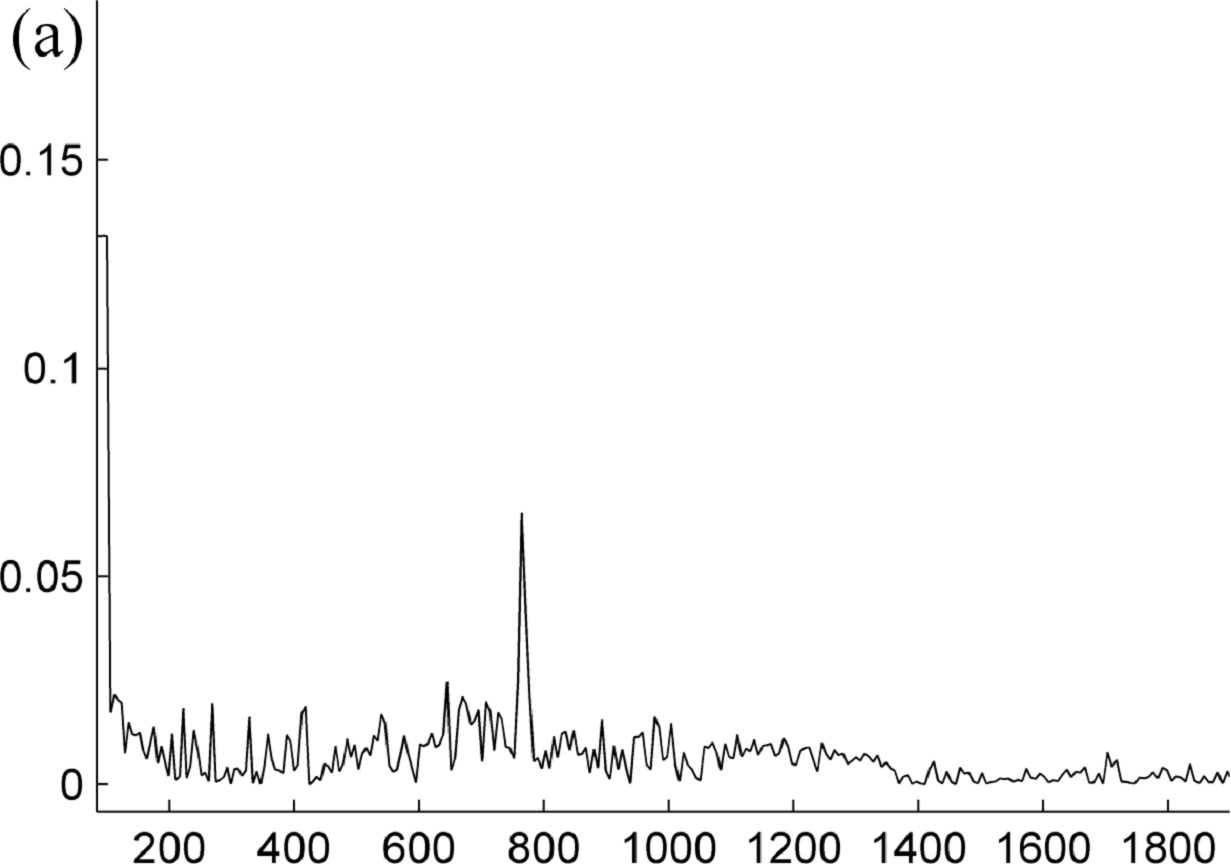
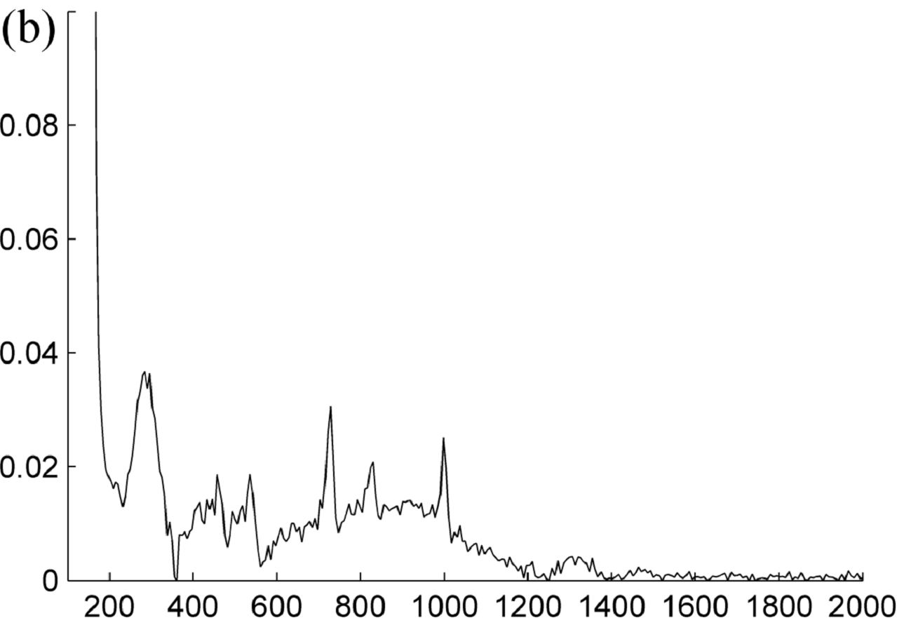
Collinear illumination of a sample simplifies the optical system and allows us to work towards a simpler more compact method to do CARS microscopy.
The recent focus of the project has been to use compressed broadband output from all normal dispersion (ANDi) photonic crystal fibre (PCF) for nonlinear microscopy applications. The preparation for using this light in nonlinear microscopy is now complete and more focus will shift to this application in future.
Research visits from Prof Parker and Dr Andy Ward (Rutherford Appleton Laboratories), Dr Robert Pal (Durham University, Prof Rainer Heintzmann and Dr Jan Rothhardt (Friedrich Schiller University), in November contributed to developments in the project, while also contributing to the ALC sponsored workshop on Laser microscopy and imaging, arranged by the group.
This project was supported by NLC rental pool funding, PISA funding, and the Newton fund.
[1] European Conference on Lasers and Electro-Optics June 2017 (CLEO-Europe): “Time-domain ptychography, reconstructing temporal objects", Dirk-Mathys Spangenberg, Michael Brügmann, Erich Rohwer, Thomas Feurer
Raman spectroscopy
Pieter Neethling
During 2012 we re-commissioned an old high resolution SPEX double monochromator, and converted it into a high resolution Raman spectrometer by fitting it with a modern intensified CCD camera as detector and using an Ar-ion laser as excitation source. This was the start of Raman activities at the LRI. In 2014 we constructed a second smaller Raman spectroscopy setup, significantly improving the experimental acquisition times and signal to noise levels. This improved our capabilities significantly and allowed us to accept an MSc student onto the project.
At the beginning of 2014, Cathrine Pfukwa joined the project as an MSc student. She started looking at the applicability of Surface Enhanced Raman Spectroscopy (SERS) for identifying and characterising important bio-molecules such as carbohydrates and amino-acids.
During 2015 she improved the synthesis of silver nanoparticles using two different protocols, as well as the synthesis of gold nanoparticles. She completed a large set of concentrations studies on the amino-acids, focussing on the lowest detection limit achievable using SERS. She graduated in March 2016. Fortunately she decided to stay on as a PhD student and spent half of 2016 at the Rutherford Appleton Laboratories in the UK as part of our collaboration with Prof Tony Parker, funded through the SA-UK Newton Fund. She is examining structural changes in anti-microbial peptides using Surface Enhanced Raman Spectroscopy and complementary vibrational spectroscopic techniques, with the aim of correlating these structural changes to the bio-activity of the peptides. These peptides form different conformational aggregates on surfaces, depending on the surface and the peptides environment. SERS therefore provides an ideal tool to investigate these conformations as it is sensitive to surface interactions. This project also forms part of a collaboration with Prof Marina Rautenbach from our Biochemistry department. She is a world expert on anti-microbial peptides and is providing us with the samples as well as the relevant biochemical analysis.
Pieter Neethling visited the Rutherford Appleton Laboratories for a week during 2017, as part of the ongoing Newton fund collaboration. During this visit, preparations were made to apply for access time to the LifeTime facility at RAL for performing two-dimensional infrared spectroscopy on the antimicrobial peptides. This application was successful and the experiments are planned for February 2018. Pieter will again visit RAL during this time to take part in the measurements. This work has also fostered a new collaboration with Prof Neil Hunt from the University of Strathclyde, who is an expert on two-dimensional infrared spectroscopy. Cathrine is progressing well and it is expected that she will finish her PhD at the end of 2018 or early 2019.
Laser techniques and theory in biological systems
Kristian Müller-Nedebock
With colleagues (Prof A. Parker, Prof S. Botchway, Dr A. Ward and Dr J. Bernardino de la Serna) at the Central Laser Facility at the Rutherford Appleton Laboratory in Oxfordshire, we started to synthesise giant unilamellar vesicles and grow tubulin filaments in confining geometries. These can be viewed as model biological systems that can be probed for their physical properties, and specifically how confinement influences cytoskeletal structure. Funding came from a Newton Fund UK-SA project through the Laser Research Institute in Stellenbosch. The experiments were done during January 2017, and January 2018, entailing synthesis and imaging of such systems, through confocal, super-resolution and light-sheet microscopy. The results will be compared with theoretical predictions for these situations.
Super Resolution Microscopy
Dr Gurthwin Bosman, Prof EG Rohwer
This project focusses on developing imaging systems to locate and track fluorescent objects far below the optical diffraction limit. The systems will initially investigate diffusional mechanisms in novel thin polymer films and the intracellular environment.
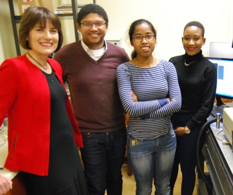
| Photo taken to celebrate the installation of a new scientific grade camera. From left: Prof Louise Warnich (Dean of Science), Dr Gurthwin Bosman (principle investigator), Miss Ratsimandresy Miora and Miss Charmaine Sibanda |
During 2017 the primary objectives; to examine single object diffusion in polymers thin films and to incorporate point spread function engineering using phase control of light in order to obtain orientation information of single dipole emitters were successfully achieved. The two objectives incorporated careful sample preparation, proper optical alignment, proficient data collection, image processing and initial model verification. The two MSc students involved in the project, Miss Ratsimandresy Holinirina Dina Miora and Miss Charmaine Sibanda were instrumental in the current successes of the project.
The students also presented their work at the local annual South African Institute of Physics conference hosted at Stellenbosch University by the Physics Department. The quality of their contributions were well received. This was verified with Miss Sibanda receiving the award for “Most Promising Young Scientist" in the Photonics Division. In addition Miss Sibanda also presented her work abroad at the OSA-JSAP symposium in Fukuou Japan, September 2017.
In 2018 greater focus will be placed on demonstrating the benefits of the current imaging system to in vivo applications. Herein the project receives guidance from local (Prof K Muller-Nedebock, SU Physics) and international (Dr Robert Pal, Durham University, UK) collaborators. Furthermore financial support for 2018-2019 has already been secured from the CSIR NLC rental pool program.
Entomological LIDAR
Erich Rohwer, Pieter Neethling
We have a collaboration on entomological LIDAR in collaboration with Dr Mikkel Brydegard and Alem Gebru at the University of Lund. The project entails the monitoring of atmospheric insects using either passive (sunlight and diffuse scattered surface light) or active (laser) light. During 2017 a publication resulted from the collaboration, but there was no experimental activity at the LRI.
